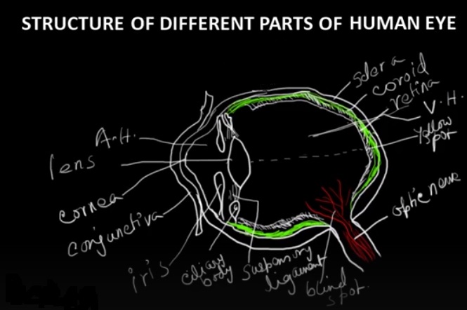
The human eye is a highly specialized sense organ that allows us to see and interpret our surroundings visually. It consists of several distinct parts, each with its own unique structure and function. Understanding the structure and functions of these different components is crucial for gaining knowledge about how our vision works. Let's take a closer look at the different components of the human eye::
Cornea:
The cornea is the clear, curved front surface that serves as the outermost layer of the eye. It acts as a protective covering and is responsible for refracting or bending light as it enters the eye. The cornea contributes significantly to focusing light onto the retina, aiding in clear vision. It also plays a crucial role in protecting the inner structures of the eye from dust, debris, and foreign objects.
Iris:
The iris is the pigmented, circular part of the eye located behind the cornea. The iris contains muscles that control the pupil, which is the central opening of the eye. These muscles either tighten or loosen to change the pupil's size, helping to manage how much light enters the eye. This process, known as pupillary reflex, helps in controlling the intensity of light reaching the retina.
Pupil:
Situated behind the iris, there exists a flexible and transparent structure known as the lens. It serves as the entry point for light into the eye. When exposed to bright light, the pupil narrows to limit the light intake and protect the delicate retina from potential harm. In low light conditions, the pupil dilates or becomes larger, allowing more light to enter, enhancing visual sensitivity.
Lens:
Positioned just behind the iris, the lens is a clear and flexible structure that helps focus light precisely onto the retina. It changes its shape in a process known as accommodation, allowing the eye to adjust focus for objects that are near or far. This shape-shifting ability is controlled by the ciliary muscles that surround the lens. When the ciliary muscles contract, the lens thickens, increasing its refractive power for near vision. On the other hand, when the ciliary muscles loosen, the lens flattens, decreasing its ability to bend light, which helps the eye focus on distant objects.
Retina:
The retina is a thin, light-sensitive layer of tissue that lines the back of the eye. It contains millions of specialized cells called photoreceptors, which convert light into electrical signals. In the retina, there exist two distinct kinds of light-sensitive cells: rods and cones. Rods are more numerous and are primarily responsible for vision in low light conditions. Rod cells don't detect color but are essential for peripheral vision and sensing movement. In contrast, cone cells are fewer in number and are mainly found in the central part of the retina, known as the fovea. Cones enable us to see colors, focus sharply, and notice fine visual details.
Optic Nerve:
The optic nerve is made up of more than a million nerve fibers and is responsible for transmitting visual signals from the retina to the brain. It serves as the main communication pathway between the eye and the brain's visual centers. At the point where the optic nerve leaves the eye, there is a small area called the optic disc or blind spot. This is where the nerve fibers exit the eye, and there are no photoreceptors. The brain fills in the missing information, so we don't perceive a gap in our visual field.
Vitreous Humor:
The space between the lens and the retina is filled with a clear, jelly-like material known as the vitreous humor. It helps preserve the eye’s round shape and supports its internal components. Additionally, it aids in directing light toward the retina and helps keep the retina firmly in place against the inner wall of the eye.
Sclera:
The outer layer of the eye, known as the sclera, is a resilient and fibrous structure. It surrounds the majority of the eye’s outer surface, giving it the characteristic white color. The sclera acts as a protective layer and provides firmness and shape to the eye. It helps maintain the shape of the eyeball and serves as an attachment site for the eye muscles that control eye movement.
 Facebook
 Twitter
 Linkedin